 |
 |


- Search
| Int Neurourol J > Volume 22(1); 2018 > Article |
|
ABSTRACT
Purpose
To characterize the electromyographic activity of abdominal striated muscles during micturition in urethane-anesthetized female mice, and to quantitatively evaluate the contribution of abdominal responses to efficient voiding.
Methods
Cystometric and multichannel electromyographic recordings were integrated to enable a comprehensive evaluation during micturition in urethane-anesthetized female mice. Four major abdominal muscle domains were evaluated: the external oblique, internal oblique, and superior and inferior rectus abdominis. To further characterize the functionality of the abdominal muscles, pancuronium bromide (25 μg/mL or 50 μg/mL, abdominal surface) was applied as a blocking agent of neuromuscular junctions.
Results
We observed a robust activation of the abdominal muscles during voiding, with a consistent onset/offset concomitant with the bladder pressure threshold. Pancuronium was effective, in a dose-dependent fashion, for partial and complete blockage of abdominal activity. Electromyographic discharges during voiding were significantly inhibited by applying pancuronium. Decreased cystometric parameters were recorded, including the peak pressure, pressure threshold, intercontractile interval, and voiding duration, suggesting that the voiding efficiency was significantly compromised by abdominal muscle relaxation.
Conclusions
The relevance of the abdominal striated musculature for micturition has remained a topic of debate in human physiology. Although the study was performed on anesthetized mice, these results support the existence of synergistic abdominal electromyographic activity facilitating voiding in anesthetized mice. Further, our study presents a rodent model that can be used for future investigations into micturition-related abdominal activity.
Micturition requires the participation of subtle sensorimotor regulatory mechanisms for the urinary bladder to be efficiently emptied when socially and behaviorally appropriate. At the nerve-control level, micturition cycles of the bladder filling with urine followed by voiding events requires systematic cooperation among afferent, somatic, and efferent nervous pathways, with efferent pathways stemming from both the central and the autonomic nervous system [1]. Similarly, the muscle-controlled component involves contraction of the bladder detrusor, relaxation of the urethra and the external urethral sphincter, and participation of additional pelvic and perineal striated muscles [2,3]. Other striated muscle regions, especially from the perineal and abdominal muscles, have also been reported to exhibit synergistic activation patterns that could facilitate urination [2,4]. Despite these arguments, the concurrent activation of the abdominal musculature during micturition has remained a controversial topic in the physiology of urination that requires further investigation.
Preclinical studies evaluating the activation of abdominal muscles during micturition using electromyographic (EMG) approaches in rats have provided further information about the physiological requirement of abdominal-visceromotor activity for facilitating voiding events in rodents [5,6]. These observations have broadened our understanding of the voiding reflex, and have raised potential voiding dysfunction issues regarding abdominal neurectomy and postoperative residual curarization that can often result in either paralysis of abdominal muscle groups or loss of this reflex activity after spinal cord injury [7-9]. Therefore, it is necessary to critically assess, in well-standardized rodent models, the contribution of the abdominal muscles to efficient voiding contractions.
By integrating the use of open cystometry (CMG) and a multielectrode EMG system in urethane-anesthetized female mice, we determined whether concomitant activation of abdominal muscles with detrusor contractions occurred during voiding. Secondly, we attempted to quantitatively determine the functionality of the abdominal muscles during micturition, and finally, we investigated the contribution of the abdominal musculature to functional and effective voiding events by blocking nicotinic-type acetylcholine receptors. Although the anesthetic plane may have affected the neurological control of the lower urinary tract, the results of this study could be useful for better understanding the physiology of micturition in rodents, and for determining the effectiveness of new treatments aimed at improving micturition.
All procedures performed in this study were approved by our Institutional Animal Care and Use Committee (IACUC; ID: AUP-0615-0044/IS00001252), and performed in accordance with the guidelines from the National Institutes of Health on the care and use of laboratory animals [10].
A total of 6 female C57BL6/J mice (24 to 26 weeks old) were tested in this study. Animals were housed in a pathogen-free environment, with 12-hour light/dark cycles, a controlled room temperature of 25˚C, and ad libitum access to food and water in plastic cages containing corncob bedding.
General anesthesia was induced after bilateral subcutaneous injections of urethane (1.2 g/kg) in the thoracic area. Although the voiding responses in urethane-anesthetized animals are not comparable to those seen in awake animals, this anesthetic helps to maintain functional responsiveness of the spinal micturition reflex in rodents [11]. After reaching the anesthetic plane (about 45 minutes), a small abdominal incision was carefully performed to expose the urinary bladder, with special care taken to minimize tissue damage and to keep the rectus abdominis muscle as intact as possible (<1-cm incision). A suprapubic catheter (PE-50) was placed into the bladder dome and fixed with a 6-0 surgical suture. The abdomen incision was then sutured (5-0 silk suture) while leaving the skin flaps open. The bladder catheter was connected to a 3-way valve that was used for the intravesical infusion of saline via an infusion pump, and for the simultaneous measurement of bladder pressure using a transbridge amplifier (World Precision Instruments, Sarasota, FL, USA). After verifying the absence of leaks, the urinary bladder was fully emptied before starting each experiment.
Eight custom-made stainless steel spring electrodes (internal diameter, 0.457 mm) were placed on different muscle surfaces to obtain bilateral EMG recordings from 4 different abdominal muscle domains: the external oblique (EO), internal oblique (IO), and the superior and inferior regions of the rectus abdominis (sRA/iRA) muscles, as shown in Fig. 1A [12]. The placement of all electrodes was verified with a stereomicroscope before initiating the CMG/EMG evaluations. An additional spring electrode was positioned on the chest, in proximity to the heart region, to make electrocardiogram (ECG) recordings and to monitor the vital condition of each animal.
Both EMG and ECG recordings were amplified with a RHD2000 amplifier (Intan Technologies, Los Angeles, CA, USA) at a sampling rate of 20 kHz and gain of 200. Signals for CMG were simultaneously recorded at 5 kHz using an auxiliary input port on the amplifier, where the transbridge amplifier was connected. All electrical and cystometric signals were acquired during constant saline solution (0.9% NaCl) infusion at rate of 20 μL/min.
After bladder filling with saline to trigger a first voiding, 5 additional voiding events were recorded as described above. Then, without stopping the infusion pump, a solution of pancuronium bromide (Sigma-Aldrich, St. Louis, MO, USA) was carefully applied to the surface of each muscular region where recording was being conducted. Using a micropipette, a total volume of 50 μL of pancuronium solution per abdominal side was applied on each muscle region. An initial concentration of 25 μg/mL (the Panc-25 group), followed by 50 μg/mL pancuronium (the Panc-50 group) was applied successively for partial and complete blockage of muscle activity, respectively. No washouts were performed between drug applications. Five additional voiding events were recorded after each application of the neuromuscular blocking agent. Values were acquired and compared at 3 different stages: intact (only saline infusion), and after the application of 25- and 50-μg/mL pancuronium. One animal died shortly after the application of 50-μg/mL pancuronium, so only 1 voiding event was recorded and included in the group analysis. In this case, we suspect that the highly concentrated pancuronium solution perfused into the abdominal cavity, causing rapid cardiac arrest. At the end of the evaluations, the animals were euthanized with CO2 while remaining under deep urethane anesthesia.
All EMG and CMG data were simultaneously acquired using an Intan interface software and saved for offline signal processing, as described below. Briefly, the CMG signals were downsampled to 62.5 Hz and smoothed with a 15-data-point window length to remove the high-frequency components using MatLab (MathWorks, Natick, MA, USA). Because the animals were in a supine position for electrode placement, the cystometric parameters (Fig. 1B) were calculated from the pressure profile and included the peak pressure for bladder contractions (cm H2O), intercontractile intervals (seconds), pressure threshold (cm H2O), voiding duration (seconds), and pressure drop slope (cm H2O/sec; Fig. 1C). These values were determined in an automated manner using custom-made MatLab scripts. All voiding events were analyzed for the intact conditions and after the application of 25- or 50-μg/mL pancuronium. The EMG and ECG signals were down-sampled to 5 kHz, and comb-filtered at 60, 120, 180, 240, and 300 Hz to remove powerline noise and harmonics. Thereafter, a second-order Butterworth band-pass filter from 10 to 1,000 Hz was applied. A template subtraction method was employed to remove ECG artifacts in the EMG channels [13], followed by an independent component analysis denoising technique to further secure the elimination of baseline noise and residual ECG artifacts. A threshold of 5 times the standard deviation of the baseline was used to determine both the onset and offset of abdominal muscle activation in the denoised signal [14,15]. A 1-second window was then selected for evaluating both the resting and voiding periods and for calculating the root mean square (RMS) value of each voiding event on each EMG channel (Fig. 1C). The temporal delays between the EMG onset and the CMG pressure peak, from the pressure peak to the EMG offset, and the pressure threshold for EMG onset/offset were also calculated to evaluate simultaneous temporal correlations. Because the EMG amplitudes were not symmetric on either the left or the right flank, and to reduce bias for interpretation, the data were analyzed based on the 4 muscle groups instead of the 8 recording channels.
The 2-tailed paired Student t-test was used to compare the statistical differences in the CMG and EMG parameters before and after the application of each dose of pancuronium. Values are shown as the mean±standard error of the mean for the voiding events under each condition and animal. The level of statistical significance was set as P<0.05.
Representative overlaid plots of EMG (black trace) activity and simultaneous CMG activity (red trace) during a voiding event are shown in Fig. 2. The pressure data are presented for a single peak voiding pressure, where high-frequency oscillations were observed after reaching the maximal pressure in all cases. The 4 abdominal muscles were activated concurrently with detrusor contractions during voiding events. Specifically, robust EMG discharges were recorded during micturition events for the EO (Fig. 2A), iRA (Fig. 2B), and IO (Fig. 2C) muscles, while the EMG signals remained low for the sRA (Fig. 2D). Higher timeresolution traces are shown in Fig. 2E–H. After the application of 25-μg/mL pancuronium, both resting and contracting EMG amplitude were attenuated for the EO (Fig. 2I), iRA (Fig. 2J), IO (Fig. 2K), and sRA (Fig. 2L) muscles. Further application of 50-μg/mL pancuronium eliminated EMG signals in the iRA (Fig. 2N), IO (Fig. 2O), and sRA (Fig. 2P) muscles, with residual activity in the EO muscles (Fig. 2M). Unexpectedly, clear differences in both amplitude and firing patterns were observed in the muscle-region paired electrodes.
The RMS value in skeletal muscles has been considered to be an indication of signal amplitude, and therefore associated with the strength of motor unit activity during EMG evaluations. Because the automated detection of EMG discharge onset/offset often failed to recognize minor firing events, a 1-second window after reaching the peak pressure value was selected for calculating the voiding RMS. The overall average RMS value for all 4 abdominal muscles was significantly reduced during resting conditions by the application of pancuronium independently of the concentration (Fig. 3A; P<0.05 for both Panc-25 and Panc-50 vs. intact). Moreover, the application of pancuronium on the abdominal surface reduced the average RMS value on all muscles during voiding contractions in a dose-dependent fashion (Fig. 3B; P<0.05 for Panc-25 vs. intact, and P<0.01 for Panc-50 vs. intact). Similarly, in the individual abdominal regions, we found that the RMS during voiding contractions was significantly reduced by the application of pancuronium in the EO, IO, and iRA muscle regions, but no significant changes were observed for the sRA muscle after neuromuscular action blockage (Fig. 3C).
A gradual rise in EMG amplitude during the bladder pressure increase, specifically from the pressure threshold to the peak pressure (pink shadows in Fig. 2), was observed. Automated onset detection analysis showed that the EMG responses of the EO, sRA, IO, and iRA muscles were activated at similar time points before the pressure peak was reached (Fig. 4A). Similarly, the offset times for the EO, sRA, IO, and iRA EMG recordings (green shadows in Fig. 2) were similar for all muscles with respect to the peak pressure (Fig. 4B). Although the IO showed reduced onset values, no significant differences were found in the overall onset and offset time across muscles and conditions (P>0.05). The EMG onset with respect to bladder threshold pressure was calculated as 25.05±1.35 cm H2O, whereas the offset was 23.22±0.74 cm H2O. No statistically significant differences were determined for these 2 values.
Representative CMG responses from the same mouse are shown during control perfusion (Fig. 5A), and after the application of either 25 μg/mL (Fig. 5B) or 50-μg/mL pancuronium to the abdominal muscle (Fig. 5C). Group analysis of the primary cystometric parameters in intact conditions and after the application of 25- or 50-μg/mL pancuronium are shown in Fig. 6. Values for peak bladder pressure, threshold pressure, and intercontractile intervals were only reduced after the surface application of 50-μg/mL pancuronium (Fig. 6A, B, and C, respectively). Voiding duration, calculated as the beginning and end of a bladder contractile event, was significantly reduced by either concentration of the nicotinic receptor antagonist (Figs. 5A–C and 6D). No significant differences were found for the pressure drop slope in the intact (-3.81±0.61 cm H2O/sec), Panc-25 (-3.99±0.56 cm H2O/sec), or the Panc-50 (-3.80±0.62 cm H2O/sec) groups.
Abdominal muscles participate in pelvic expulsive functions, such as defecation and labor [4,9,16,17]. The findings of the present study corroborate that abdominal muscles were also activated during micturition in a female mouse model, and demonstrate that this reflex seems to have exerted a synergistic effect on the efficiency of voiding events. Previous studies have demonstrated that abdominal visceromotor activity was maintained by the pelvic afferent sensory branch, and that activity was intensified by acetic acid through chemical-sensing receptors [5,18]. Our results suggest that abdominal muscles were reflexively activated when the intravesical pressure surpassed approximately 25 cm H2O, with an overall duration of 3 to 5 seconds. As a result, the relatively consistent onset pressure may also suggest that motor neurons innervating the abdominal muscles are recruited through bladder mechanoreceptors when bladder pressure is increased. Previous studies in humans have reported that during voluntary contractions, an initial onset of EO is seen, followed by the pelvic floor and RA muscles, and finally by IO activity [19]. However, our results show varying, but not significantly different, onset and offset times in reference to the pressure peak for the EO, IO, and iRA muscle responses. The difference in the sRA activation period is presumably due to the low EMG amplitude recorded, as shown in Fig. 2B. Although activation can be observed in sRA muscle recordings, the low signal-to-noise ratio greatly impaired the distinguishability of activation periods from resting stages. These results may be considered as further evidence that the abdominal activity during voiding is a reflex event, and different from voluntary contractions, although the physiological differences between human and rodent models need to be considered for an integrative interpretation.
Abdominal muscle straining during voiding in humans is usually considered to be a symptom of bladder outflow obstruction [20,21]. However, clinical observations have failed to demonstrate a consistent relationship between abdominal straining and micturition in healthy subjects or in those with bladder outlet obstruction, suggesting the possibility of nonpathological muscle activity [4,22,23]. The maintenance of abdominal pressure has also been later proven to be an important factor in promoting vesical contraction in humans [24], as well as in animal investigations that confirmed the function of abdominal musculature in improving voiding efficiency [5,6]. For example, it was suggested that contraction of abdominal muscles also contributed to the activation of the pelvic musculature, especially the levator ani muscle, which consequently affected the maintenance of urethral pressure during voiding [4,5]. Similarly, preclinical animal models discovered concurrent discharges of abdominal muscles in both awake and anesthetized female rats, and further investigated the role of sensory pathways in modulating abdominal muscle activity [2]. Based on these studies, a distinct role for the activity of nociceptive abdominal visceromotor responses from that of voiding-associated abdominal responses has been proposed in female rats [6,22], and the authors believed abdominal activity to be important for maintaining high-frequency oscillations as a key feature of voiding patterns in anesthetized rats. Supporting these observations, the recording of maximal intra-abdominal pressure during voiding onset reinforced the necessity of constant abdominal musculature straining for voiding events in awake rats [25].
Altogether, our results and those of the reports discussed above further the idea that abdominal muscles are required for micturition in rodents, as ‘chemical silencing’ with pancuronium significantly affected voiding efficiency. In addition to its supportive role in facilitating urination, a seemingly contradictory function of abdominal straining in maintaining urinary continence has been reported [26,27]. The specific contraction pattern of abdominal muscles, as well as the coactivation of pelvic floor muscles, can contribute to controlling urine flow by, on the contrary, increasing urethral pressure [28,29]. These results have been supported by studies of various therapies involving abdominal muscle training for preventing and treating urinary incontinence [27]. Nonetheless, how the coactivation of 2 muscle groups has different outcomes in reflexive and voluntary activity requires further exploration.
Previous studies evaluating the functional effects of the neuromodulation of pathways innervated by the pelvic nerve on the pelvic musculature have suggested that abdominal muscles play a role in the micturition process [5,18]. Based on bilateral innervation by the pelvic nerve, as well as the bilateral placement of the recording electrodes, a symmetric EMG pattern was expected for each recording region in the mouse abdomen. However, we did not find that to be the case because clear distinctions in amplitude and firing patterns were detected in the muscle-region paired electrodes. We speculate this could have been caused by various factors that are hard to control experimentally, such as differences due to the manual placement of electrode locations and variations in the impedance of the tissue-electrode interface, the intramuscular content of abdominal fat, and cholinergic innervation. Therefore, to reduce bias in data interpretation, our results were analyzed based on 4 muscle groups instead of 8 recording channels. Based on the above considerations, it can be suggested that the multiple-electrode placement used in this study may be an important technique for obtaining more comprehensive information, especially when studying isolated muscular activity in abdominal muscles.
The CMG evaluations during saline infusion suggest that the abdomen plays a significant role in voiding; nonetheless, the targeted blockage of abdominal muscular activity further provided a more specific understanding of the role of abdominal visceromotor activity in facilitating micturition. Our study suggests that local paralysis of the neuromuscular junction is a very useful tool when evaluating abdominal muscle physiology. A parallel approach using botulinum toxin A to induce paralysis of the rectus abdominis muscle resulted in compromised voiding efficiency with decreased voided volume and average urine flow [6]. However, because both the EO and the IO muscles discharge similarly and intensively during voiding, simple blockage of the rectus abdominis is not enough to provide evidence supporting the importance of the full abdomen for micturition.
Pancuronium bromide has been employed in multiple studies as a neuromuscular blocking drug that prevents the activation of acetylcholine receptors [30-32]. However, as pancuronium may also impact the activation of muscarinic receptors in smooth muscle, we decided to apply it onto the abdominal surface instead of intravenously to avoid negative effects on the bladder, heart, and respiratory function [33,34]. The concentrations of the applied pancuronium were selected after preliminary testing in 3 mice that were not included in this report. We found that anesthetized female mice could maintain consistent voiding patterns at a pancuronium concentration of 25 μg/mL for more than 20 minutes, but after application of a pancuronium solution of 50 μg/mL, most of the muscular activity in the recorded abdominal regions was eliminated.
In this study, the chemically-induced paralysis of the abdominal musculature reduced the intravesical pressure peak by 18.9%, the intercontractile interval by 47.8%, the pressure threshold by 50.5%, and the voiding duration by 23.2%, suggesting that voiding efficiency was significantly compromised when abdominal muscles did not participate in the micturition process. As the maximal bladder pressure was compromised, we expected a reduced value of the pressure drop slope. However, no significant differences were found for this specific parameter. These results quantitatively support the importance of the reflexive activation of the abdominal musculature for maintaining micturition efficiency in mice. In patients with urine retention, biofeedback of the pelvic floor musculature is often required for maintaining continence and improving voiding efficiency. Indirectly, our results also support the beneficial effects of abdominal muscle training as a urological rehabilitation alternative that needs to be encouraged [35].
It has been considered that the contraction of the abdominal muscles contributes to the increase in intra-abdominal pressure, consequently affecting the intravesical pressure [6]. Unfortunately, intra-abdominal and urethral pressures were not recorded in this study, which somewhat limited the degree to which we could explore in detail the mechanisms of abdominal muscle discharge. Meanwhile, the voided volume was not measured in this study, so compromised voiding efficiency could only be revealed by indirect evidence, such as decreased peak pressure, intercontractile interval, and voiding duration. Direct measurement of the voided volume would provide more details for helping us to interpret the functionality of abdominal muscles in micturition.
Nonvoiding contractions were observed during bladder filling in 2 of the animals that were tested (not shown). Although the female mice used in this experiment were ~6 months old, no age-related detrusor overactivity was expected. Because of this situation, we believe that the increased number of nonvoiding contractions may have been related to the placement of the suprapubic catheter, which perhaps damaged the sensory nervous axons. Although the EMG activity did not show a close correlation with the nonvoiding bladder pressure fluctuations, it is a very interesting observation that an increased baseline RMS value was found in these animals, further suggesting the plausibility of an association of abdominal muscle activation with bladder overactivity. Whether these EMG findings of the abdominal muscles are reproducible in humans, or can be used as a marker for overactive bladder diagnosis and prognosis, remains a topic for future experimental evaluations in healthy patients and those with urological symptoms.
Our results support the presence of a synergistic role for abdominal muscle activation during micturition in anesthetized female mice. The targeted blockage of abdominal muscles, including the EO, IO, sRA, and iRA, proved that they exert a significant function in maintaining voiding efficiency. This study provides an animal model for comprehensively investigating abdominal muscle functions under pathological conditions. Despite the limitations of the present study, our results will advance our understanding of visceromotor activity in the abdominal muscles during voiding.
NOTES
Grant/Fund Support
This work was supported by NIH DK082644 (YZ), the University of Houston (YZ), the Brown Foundation (TBB, AM), and the Houston Methodist Foundation (TBB, AM).
Research Ethics
All procedures performed in this study were approved by our Institutional Animal Care and Use Committee (IACUC; ID: AUP-0615-0044 / IS00001252. Responsible investigator: Alvaro Munoz), and performed in accordance with the guidelines from the National Institutes of Health on the care and use of laboratory animals.
AUTHOR CONTRIBUTION STATEMENT
·Full access to all the data in the study and takes responsibility for the integrity of the data and the accuracy of the data analysis: H Choi
·Study concept and design: JH Bae
·Acquisition of data: C Zhang, A Munoz
·Analysis and interpretation of data: C Zhang, Y Zhang, Y Cruz, TB Boone, A Munoz
·Drafting of the manuscript: C Zhang, A Munoz
·Critical revision of the manuscript for important intellectual content: C Zhang, Y Zhang, Y Cruz, TB Boone, A Munoz
·Statistical analysis: C Zhang, A Munoz
·Obtained funding: Y Zhang, A Munoz, TB Boone
·Study supervision: A Munoz
ACKNOWLEDGEMENTS
The authors would like to thank Dr. Betsy Salazar and Mr. Kris Hoffman for helping to set up the electromyography system.
REFERENCES
1. Fowler CJ, Griffiths D, de Groat WC. The neural control of micturition. Nat Rev Neurosci 2008;9:453-66. PMID: 18490916


2. Cruz Y, Downie JW. Sexually dimorphic micturition in rats: relationship of perineal muscle activity to voiding pattern. Am J Physiol Regul Integr Comp Physiol 2005;289:R1307-18. PMID: 15994373


3. Corona-Quintanilla DL, Zempoalteca R, Arteaga L, Castelán F, Martínez-Gómez M. The role of pelvic and perineal striated muscles in urethral function during micturition in female rabbits. Neurourol Urodyn 2014;33:455-60. PMID: 23636895


5. Cruz Y, Downie JW. Abdominal muscle activity during voiding in female rats with normal or irritated bladder. Am J Physiol Regul Integr Comp Physiol 2006;290:R1436-45. PMID: 16373437


6. Smith PP, Smith CP, Boone TB, Somogyi GT. Is abdominal wall contraction important for normal voiding in the female rat? BMC Urol 2007;7:5. PMID: 17343732


7. de Lima E Souza R, Apgaua BT, Milhomens JD, Albuquerque FT, Carneiro LA, Mendes MH, et al. Severe fat embolism in perioperative abdominal liposuction and fat grafting. Braz J Anesthesiol 2016;66:324-8. PMID: 27108833


8. Estenne M, Pinet C, De Troyer A. Abdominal muscle strength in patients with tetraplegia. Am J Respir Crit Care Med 2000;161(3 Pt 1):707-12. PMID: 10712311


9. Hascakova-Bartova R, Dinant JF, Parent A, Ventura M. Neuromuscular electrical stimulation of completely paralyzed abdominal muscles in spinal cord-injured patients: a pilot study. Spinal Cord 2008;46:445-50. PMID: 18227852


10. National Research Council (U.S.). Committee for the Update of the Guide for the Care and Use of Laboratory Animals., Institute for Laboratory Animal Research (U.S.), and National Academies Press (U.S.), Guide for the care and use of laboratory animals. 8th ed. Washington, D.C.: National Academies Press; 2011.
11. Andersson KE, Soler R, Füllhase C. Rodent models for urodynamic investigation. Neurourol Urodyn 2011;30:636-46. PMID: 21661007


12. Salazar BH, Hoffman KA, Zhang C, Kavanagh A, Zhang Y, Boone TB, et al. Electrical activity of the bladder is attenuated by intravesical inhibition of P2X2/3 receptors during micturition in female rats. Int Neurourol J 2017;21:259-69. PMID: 29298472


13. Drake JD, Callaghan JP. Elimination of electrocardiogram contamination from electromyogram signals: an evaluation of currently used removal techniques. J Electromyogr Kinesiol 2006;16:175-87. PMID: 16139521


14. Li X, Zhou P, Aruin AS. Teager-Kaiser energy operation of surface EMG improves muscle activity onset detection. Ann Biomed Eng 2007;35:1532-8. PMID: 17473984


15. Solnik S, DeVita P, Rider P, Long B, Hortobágyi T. Teager-Kaiser Operator improves the accuracy of EMG onset detection independent of signal-to-noise ratio. Acta Bioeng Biomech 2008;10:65-8. 
16. Koutsomanis D, Lennard-Jones JE, Roy AJ, Kamm MA. Controlled randomised trial of visual biofeedback versus muscle training without a visual display for intractable constipation. Gut 1995;37:95-9. PMID: 7672690


17. Rankin G, Stokes M, Newham DJ. Abdominal muscle size and symmetry in normal subjects. Muscle Nerve 2006;34:320-6. PMID: 16775833


18. Cueva-Rolón R, Gómez LE, Komisaruk BR, Muñoz-Martínez EJ. Inhibition of withdrawal responses by pelvic nerve electrical stimulation. Brain Res 1995;679:267-73. PMID: 7633887


19. Madill SJ, McLean L. Quantification of abdominal and pelvic floor muscle synergies in response to voluntary pelvic floor muscle contractions. J Electromyogr Kinesiol 2008;18:955-64. PMID: 17646112


20. Reynard JM, Peters TJ, Lamond E, Abrams P. The significance of abdominal straining in men with lower urinary tract symptoms. Br J Urol 1995;75:148-53. PMID: 7531591


21. Hald T, Nordling J, Andersen JT, Bilde T, Meyhoff HH, Walter S. A patient weighted symptom score system in the evaluation of uncomplicated benign prostatic hyperplasia. Scand J Urol Nephrol Suppl 1991;138:59-62. PMID: 1723812


22. Bors E, Blinn KA. Abdominal electromyography during micturition. Calif Med 1965;102:17-22. PMID: 14243851


23. Yang JM, Huang WC. Factors associated with voiding function in women with lower urinary tract symptoms: a mathematic model explanation. Neurourol Urodyn 2003;22:574-81. PMID: 12951666


24. Shafik A. The anterolateral abdominal wall muscles during vesical filling and evacuation: electromyographic study. Urology 2008;71:621-4. PMID: 18313114


25. Lee T, Andersson KE, Streng T, Hedlund P. Simultaneous registration of intraabdominal and intravesical pressures during cystometry in conscious rats: effects of bladder outlet obstruction and intravesical PGE2. Neurourol Urodyn 2008;27:88-95. PMID: 17565725


26. Ferla L, Darski C, Paiva LL, Sbruzzi G, Vieira A. Synergism between abdominal and pelvic floor muscles in healthy women: a systematic review of observational studies. Fisioter Mov. 2016. 29:399-410. 

27. Sapsford RR, Hodges PW. Contraction of the pelvic floor muscles during abdominal maneuvers. Arch Phys Med Rehabil 2001;82:1081-8. PMID: 11494188


28. Sapsford RR, Clarke B, Hodges PW. The effect of abdominal and pelvic floor muscle activation patterns on urethral pressure. World J Urol 2013;31:639-44. PMID: 23192396


29. Sapsford RR, Hodges PW. The effect of abdominal and pelvic floor muscle activation on urine flow in women. Int Urogynecol J 2012;23:1225-30. PMID: 22278713


30. Hutter J, Habler O, Kleen M, Tiede M, Podtschaske A, Kemming G, et al. Effect of acute normovolemic hemodilution on distribution of blood flow and tissue oxygenation in dog skeletal muscle. J Appl Physiol (1985) 1999;86:860-6. PMID: 10066697


31. Löwenick CV, Krampfl K, Schneck H, Kochs E, Bufler J. Open channel and competitive block of nicotinic receptors by pancuronium and atracurium. Eur J Pharmacol 2001;413:31-5. PMID: 11173060


32. Ness TJ, Gebhart GF. Colorectal distension as a noxious visceral stimulus: physiologic and pharmacologic characterization of pseudaffective reflexes in the rat. Brain Res 1988;450:153-69. PMID: 3401708


33. Nagtegaal JE, Siero HL, Dirksen R, Lammers JW, Rodrigues de Miranda JF, Russel FG. Pancuronium masks the prejunctional muscarinic autoreceptor in guinea pig tracheal smooth muscle. Life Sci 1995;57:2325-33. PMID: 7491091


34. Wiklund NP, Ohlén A, Wiklund CU, Cederqvist B, Hedqvist P, Gustafsson LE. Neuromuscular actions of endothelin on smooth, cardiac and skeletal muscle from guinea-pig, rat and rabbit. Acta Physiol Scand 1989;137:399-407. PMID: 2556891


35. Dumoulin C, Lemieux MC, Bourbonnais D, Gravel D, Bravo G, Morin M. Physiotherapy for persistent postnatal stress urinary incontinence: a randomized controlled trial. Obstet Gynecol 2004;104:504-10. PMID: 15339760


Fig. 1.
Experimental approach and the parameters that were evaluated. (A) Diagram depicting the experimental configuration in the supine position for electromyography/cystometry (EMG/CMG): 1 suprapubic catheter at the bladder dome, 8 recording electrodes on the abdominal muscles: EO (1–2), sRA (3–4), IO (5–6), and iRA (7–8), and 1 chest electrode for electrocardiography. (B) Parameters evaluated for CMG data. (C) Parameters evaluated for EMG data. The red trace represents CMG recordings, while the gray line indicates the EMG response. Voiding duration (VD) was calculated as the time difference between the peak pressure and end of voiding. Other EMG parameters were determined as explained. EO, external oblique; IO, internal oblique; sRA, superior region of the rectus abdominis; iRA, inferior region of the rectus abdominis; PP, peak pressure; ICI, intercontractile interval; PT, pressure threshold; PDS, pressure drop slope; EV, end of voiding.
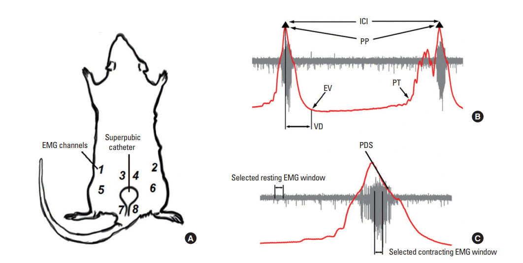
Fig. 2.
Electromyographic responses from 4 different abdominal regions of the same animal during voiding. In all panels an overlaid plot for cystometric (CMG) (red traces) and concurrent electromyographic (EMG) recordings (black traces) of the 4 evaluated muscle groups are shown. (A–D) A complete voiding event involving 4 muscle regions: (A) the external oblique (EO), (B) the superior rectus abdominis (sRA), (C) the internal oblique (IO), and (D) the inferior rectus abdominis (sRA). (E–H) A more detailed presentation of panels (A–D), respectively. The vertical line in A–H shows the mean onset (orange traces) and offset (green traces) time points of the corresponding EMG activation with respect to peak pressure, with the shaded region revealing the standard error. Panels (I–L) and (N–P) show the CMG/EMG data after successive application of 25- and 50-µg/mL pancuronium in the recording channels (A–D), respectively.
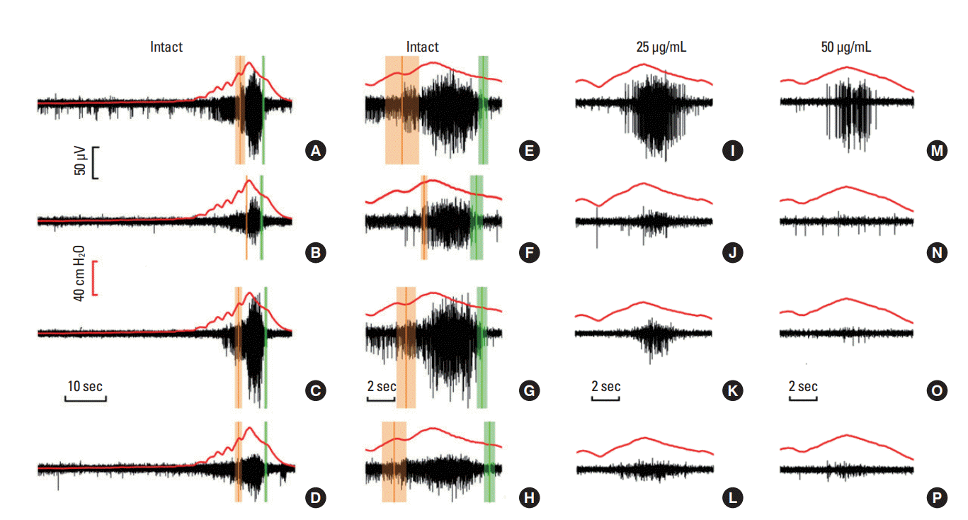
Fig. 3.
Changes in RMS as indicative of motor unit strength during voiding in 3 experimental conditions. (A, B) Comparisons between the average RMS during resting and voiding (abdominal contractions) for all 3 stages. (C) Detailed RMS values for each abdominal muscular domain studied. Intact: saline, before the application of pancuronium bromide. Application of 25-µg/mL pancuronium (Panc-25) and 50-µg/mL pancuronium (Panc-50). RMS, root mean square; EO, external oblique; sRA, superior rectus abdominis; IO, internal oblique; iRA, inferior rectus abdominis. *P<0.05 and **P<0.01 versus the corresponding intact stage, respectively.
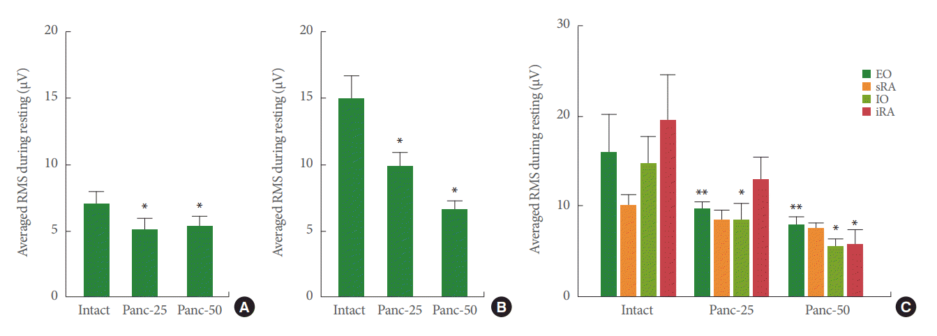
Fig. 4.
EMG onset and offset times with respect to peak bladder pressure during voiding. (A) Onset time for EMG activity in different regions of the abdominal musculature. (B) Offset time for EMG activity in different abdominal regions. EMG, electromyography; EO, external oblique; sRA, superior rectus abdominis; IO, internal oblique; iRA, inferior rectus abdominis; PP, peak pressure.
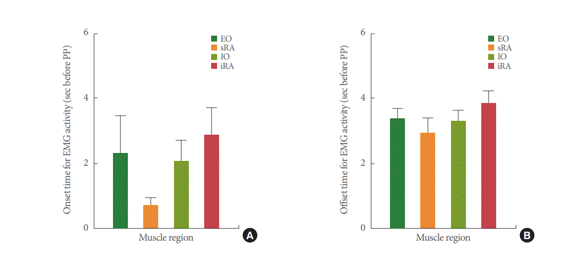
Fig. 5.
Representative cystometric responses after pancuronium application in a female mouse. (A) Bladder pressure changes during control conditions. (B) Cystometric changes induced by application of a saline solution containing 25-µg/mL pancuronium (Panc-25) to the abdominal muscles, followed by 50-µg/mL pancuronium (Panc-50). Cystometric traces are from the same mouse in a supine position, and with electromyographic electrodes placed on various abdominal regions (see Fig. 1).
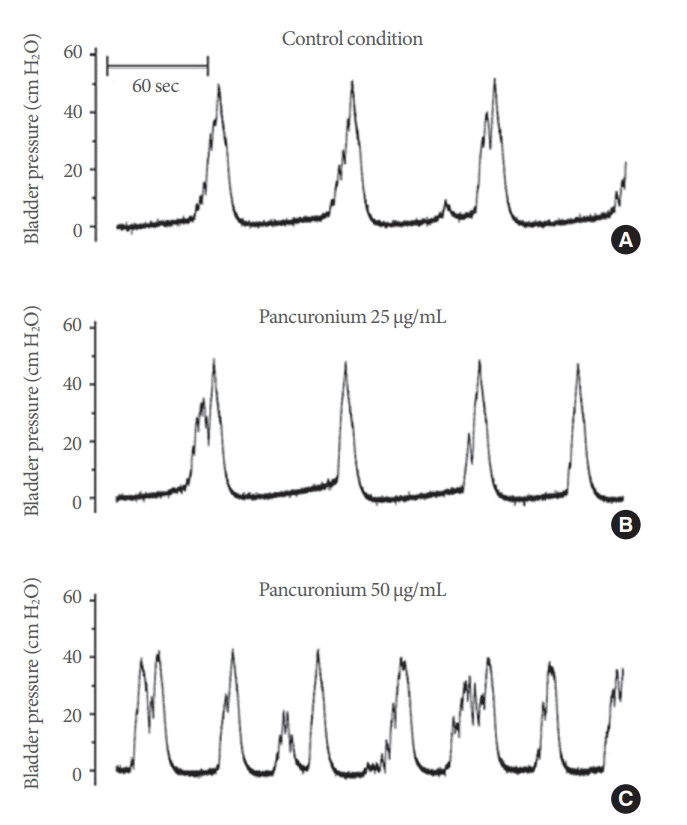
Fig. 6.
Changes on cystometric parameters after neuromuscular inhibition of abdominal muscles. (A) Peak bladder pressure. (B) Threshold pressure. (C) Intercontractile interval. (D) Voiding duration. Intact: before the application of pancuronium bromide. Panc: application of 25-µg/mL pancuronium (Panc-25) and 50-µg/mL pancuronium (Panc-50). *P<0.05 versus the corresponding intact stage.


