INJ Search

CLOSE


Int Neurourol J > Volume 25(Suppl 1); 2021 > Article
Hwang, Jin, Ko, Kim, Cho, Sung, Choi, and Chang: Polydeoxyribonucleotide Attenuates Airway Inflammation Through A2AR Signaling Pathway in PM10-Exposed Mice

ABSTRACT

Purpose

Inhalation of air containing high amounts of particular matter (PM) causes various respiratory disorders including asthma, chronic obstructive pulmonary disease, and lung cancer. The changes of expression of inflammatory factors by polydeoxyribonucleotide (PDRN) administration in the PM10-exposed trachea inflammation model were evaluated.

Methods

PM10 was administered to mouse trachea to induce acute inflammatory damage, and changes in inflammatory factors were observed after administration of PDRN and 3,7-dimethyl-1-propargylxanthine (DMPX) for 3 days daily. Expression of inflammatory cytokines, adenosine A2A receptor (A2AR), protein kinase A (PKA), 3΄,5΄-cyclic adenosine monophosphate responsive element binding protein (CREB) were detected by enzyme‐linked immunosorbent assay, immunofluorescence, and western blot assay.

Results

PM-exposed trachea showed increased tumor necrosis factor (TNF)-α and interleukin (IL)-1β expression, and expression of TNF-α and IL-1β was inhibited by PDRN treatment in PM-exposed mice. PM-exposed trachea showed increased nuclear factor (NF)-κB phosphorylation, and phosphorylation of nuclear factor-kappa B was inhibited by PDRN treatment in PM-exposed mice. PM-exposed trachea showed increased expression of A2AR, but PDRN treatment more enhanced A2AR expression in PM-exposed mice. PKA phosphorylation was not changed and CREP phosphorylation was decreased, however PDRN treatment increased phosphorylation of PKA and CREB in PM-exposed mice. DMPX treatment blocked all the effects of PDRN on PM-exposed mice, demonstrating that the action of PDRN occurs via A2AR.

Conclusions

PDRN treatment attenuated inflammation in the trachea of the PM10-exposed mice. This improving effect of PDRN can be ascribed to the activation of A2AR through the cAMP-PKA pathway.

• HIGHLIGHTS

- Expression of TNF-α and IL-1β and phosphorylation of NF-κB were inhibited by PDRN treatment.
- PDRN treatment more enhanced A2AR expression and increased phosphorylation of PKA and CREB.
- DMPX treatment blocked all the effects of PDRN on PM-exposed mice.

INTRODUCTION

Due to rapid industrialization and urbanization, air pollution in many countries is becoming increasingly severe and causing social problems such as the outbreak of respiratory diseases. Particulate matter (PM) contained in polluted air has been reported to have a detrimental effect on the human body [1]. PM is composed of various components such as polycyclic aromatic hydrocarbons and mediates toxic effects. Inhalation of air containing high amounts of PM causes various respiratory diseases such as asthma, chronic obstructive pulmonary disease, and lung cancer [2-4]. PM 10 (PM10) refers to material with a diameter of <10 μm. The small-sized PMs accumulate in the airways and lungs through breathing and affect the circulatory system [5].
Airways exposed to small-sized PMs secrete proinflammatory cytokines such as inducible nitric oxide (NO) synthase, tumor necrosis factor (TNF)-α, interleukin (IL)-1β, reactive oxygen species. Small-sized PMs directly penetrate cell membranes, causing mitochondrial membrane disruption and mitochondrial structure and function changes [6-8]. PMs-stimulated alveolar macrophages decreased cell viability and DNA function [3].
It has been reported that the increase in inflammatory factors can be reduced by adenosine A2A receptor (A2AR) stimulation in lung injury [9,10]. One of the adenosine agonists, polydeoxyribonucleotide (PDRN) is known as a candidate for anti-inflammatory drugs by inhibiting inflammation and apoptosis by selectively stimulating A2AR in lung-injured animals. PDRN is derived from salmon semen and consists of a stable mixture of nucleotides ranging in size from 50 to 1,500 kDa [10,11]. Of the 4 adenosine receptors, PDRN selectively acts on A2AR, and selective stimulation of A2AR effectively inhibits inflammatory substances [10-12].
PDRN administration increases 3΄,5΄-cyclic adenosine monophosphate (cAMP) and regulates the pathway leading to phosphorylation of protein kinase A (PKA) and cAMP responsive element binding protein (CREB) [13]. In animal model of lung injury, PDRN treatment can significantly reduce the increase in inflammatory factors through regulation of the nuclear factor (NF)-κB pathway [10]. Treatment of PDRN reduces NF-κB factor phosphorylation and the expression of inflammatory mediators such as NO, TNF-α, and interleukin-6 (IL-6). In animal model of acute lung injury, PDRN treatment can reduce apoptosis by regulating the B-cell lymphoma 2 family through activation of the cAMP pathway [14].
Although various studies have been conducted on the toxic effects of polycyclic aromatic hydrocarbons, a major component of PM, the therapeutic effect of PDRN in PM10-mediated trachea inflammation has not been identified. In this study, the changes in the expression of inflammatory factors by PDRN administration in the PM10-mediated trachea inflammation were evaluated.

MATERIALS AND METHODS

Animals and Treatments

This study was approved by the Institutional Care and Use Committee of Kyung Hee University (KHUASP [SE]-18-126). ICR male mice, 7–8 weeks old (18–20 g), were provided by the Orient Bio (Sungnam, Korea). For the enzyme‐linked immunosorbent assay (ELISA) analysis, mice were assigned into the 6 groups (n=7 in each group): sham group, PM10-exposed group, PM10-exposed with 2-mg/kg PDRN treatment group, PM10-exposed with 4-mg/kg PDRN treatment group, PM10-exposed with 8-mg/kg PDRN treatment group, PM10-exposed with 16- mg/kg PDRN treatment group. PDRN was obtained by Kyongbo Pharm. Co. (Seoul, Korea). PM10 was purchased from European Reference Materials (Retieseweg, Geel, Belgium).
For the immunofluorescence and western blot assay, mice were assigned into the 4 groups (n=7 in each group): sham group, PM10-exposed group, PM10-exposed with PDRN treatment group, PM10-exposed with PDRN and 3,7-dimethyl-1-propargylxanthine (DMPX) treatment group. DMPX were purchased from Sigma Aldrich (St. Louis, MO, USA).
Skin around neck of animals was excised to expose the trachea, and 100 μg/kg (50 μL volume) of PM10 was injected using a 1-mL syringe (30 gauge). One day after injection of PM10, mice were treated with intraperitoneal injection of 8-mg/kg PDRN or 8-mg/kg DMPX for 3 days daily.

Tissue Preparation

Animals were sacrificed 72 hours after intraperitoneal injection of PDRN. Mice were anesthetized with intraperitoneal injection of 10-mg/kg Zoletil 50 (Vibac Laboratories, Carros, France), and tracheas were collected. Trachea tissues were subsequently fixed with 4% buffered paraformaldehyde, dehydrated in ethanol gradually, infiltrated in xylene, and embedded in paraffin. The paraffin blocks were cut into 5-μm-thick coronal slices using a microtome, and the sliced tissues were placed on the gelatin-coated slides.

ELISA Assay

cAMP, TNF-α, and IL-1β concentration was obtained using enzyme immunoassay kit following the manufacturer’s manual, referring to previous paper [15]. cAMP, TNF‐α, and IL‐1β ELISA kits were purchased from Abcam (Cambridge, MA, UK). Standard and diluted samples prepared for cAMP calculation were added to each well. Labeled alkaline phosphatase-conjugate was added to each well. Then, the cAMP complete antibody was reacted for 2 hours. Stop buffer was treated and read at 405-nm wavelength (Thermo Fisher Scientific, Waltham, MA, USA). For TNF-α and IL-1β detection, standard and lysed samples were added to each well and incubated for 2 hours. Antibodies for TNF-α and IL-1β were added to each well and reacted for 1 hour. Subsequently, horseradish peroxidase (HRP)-streptavidin buffer was added and incubated for 45 minutes. In the dark, 3,3΄,5,5΄-tetramethylbenzidine development solution was added and incubated for 30 minutes. Stop buff was added to each well and read at 450 nm wavelength (Thermo Fisher Scientific).

Immunofluorescence

Immunofluorescence analysis of tracheal tissues was done, referring to previous paper [10,13]. Trachea tissues on paraffin slides were deparaffinized with xylene, rehydrated with ethanol, deionized water for 5 minutes, and denatured for 10 minutes in boiling 10mM citric acid (pH, 6.0) for 3 minutes at 100°C. Tissue slides were cooled for 30 minutes at room temperature, and blocked with 5% normal goat serum in tris-buffered saline for 1 hour. Tissue slides were reacted with primary antibody against mouse TNF-α or rabbit IL-6 in 1:200 titer overnight at 4°C. The slides were treated with secondary Alexa 488-mosue IgG or Alexa 594-rabbit IgG (1:500 dilution) antibody for 2 hours. The slides were mounted on a coverslip. Fluorescence images were acquired by Leica DMi8 fluorescence microscope (Leica Microsystems, Wetzlar, Germany) wavelength in the excitation/emission to 490/525 nm for Alexa 488 or excitation/emission to 590/617 nm for Alexa 594.

Western Blot Analysis

Western blot analysis of tracheal tissues was done by referring to previous paper [13,16]. Collected trachea tissues were homogenized in cold radioimmunoprecipitation assay buffer (RIPA buffer) with 1mM phenylmethylsulfonyl fluoride. RIPA buffer was purchased from Cell Signaling Technology (Danvers, MA, USA). Homogenized tissues were centrifuged at 14,000 g for 10 minutes at 4°C, and supernatants were collected. Protein 30 μg was separated by 15% sodium dodecyl sulfate polyacrylamide gel electrophoresis and subsequently transferred onto polyvinylidene fluoride membranes. After blocking with 5% skim milk overnight, membranes were reacted with primary antibody against mouse β-actin, A2AR, or rabbit phosphorylated PKA (p-PKA), PKA, p-CREB, CREB, p-NF-κB, NF-κB in 1:1,000 titer and then reacted with HRP-conjugated secondary antibody in 1:2,000 titer. Quantitative band intensity was evaluated using Image Pro Plus software (ver. 6; Rockville, MD, USA).

Statistical Analyses

One-way analysis of variance analysis and Duncan posttest were done using IBM SPSS Statistics ver. 23.0 (IBM Co., Armonk, NY, USA). Data were presented as mean±standard error of mean, and statistical significance was defined as P<0.05.

RESULTS

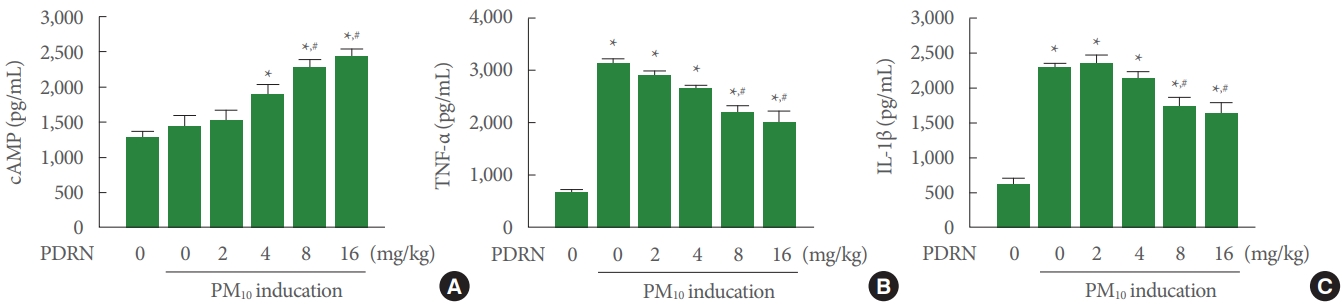
cAMP, TNF-α, and IL-1β Concentration

Fig. 1A shows that cAMP concentration was not changed in the trachea of the PM10-exposed mice. PDRN treatment increased cAMP concentration in the trachea of the PM10-exposed mice. Fig. 1B shows that TNF-α concentration was increased in the trachea of the PM10-exposed mice. PDRN treatment inhibited TNF-α concentration in the trachea of the PM10-exposed mice. Fig. 1C shows that IL-1β concentration was increased in the trachea of the PM10-exposed mice. PDRN treatment inhibited TNF-α concentration in the trachea of the PM10-exposed mice. PDRN had a significant effect at concentrations of 8 mg/kg and 16 mg/kg.

TNF-α and IL-1β Expression

TNF-α expression was increased in the trachea of the PM10-exposed mice. PDRN treatment suppressed TNF-α expression, whereas DMPX cotreatment with PDRN abolished inhibitory effect of PDRN in the trachea of the PM10-exposed mice (Fig. 2). IL-1β expression was increased in the trachea of the PM10-exposed mice. PDRN treatment inhibited IL-1β expression, whereas DMPX cotreatment with PDRN abolished inhibitory effect of PDRN in the trachea of the PM10-exposed mice (Fig. 3).

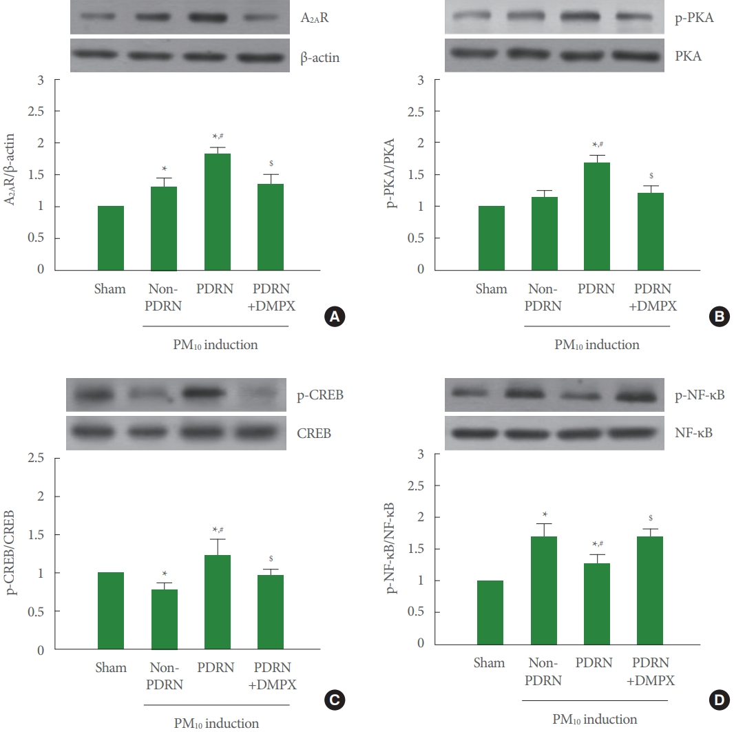
A2AR Expression and Phosphorylation of PKA, CREB, and NF-κB

Fig. 4A shows that A2AR expression was increased in the trachea of the PM10-exposed mice. PDRN treatment more increased A2AR expression, whereas DMPX cotreatment with PDRN abolished increasing effect of PDRN in the trachea of the PM10-exposed mice. Fig. 4B shows that phosphorylation of PKA was not changed in the trachea of the PM10-exposed mice. PDRN treatment increased phosphorylation of PKA, whereas DMPX cotreatment with PDRN abolished phosphorylating effect of PDRN in the trachea of the PM10-exposed mice. Fig. 4C shows that phosphorylation of CREP was decreased in the trachea of the PM10-exposed mice. PDRN treatment increased phosphorylation of CREP, whereas DMPX cotreatment with PDRN abolished phosphorylating effect of PDRN in the trachea of the PM10-exposed mice. Fig. 4D shows that phosphorylation of NF-κB was increased in the trachea of the PM10-exposed mice. PDRN treatment inhibited phosphorylation of NFκB, whereas DMPX cotreatment with PDRN abolished dephosphorylating effect of PDRN in the trachea of the PM10-exposed mice.

DISCUSSION

It is well known that PM aspiration increases inflammatory factors and accelerates apoptotic cell death [2-4,6]. Reduced cell viability and production of TNF‐α, IL‐1β, and IL‐6 were observed after exposure to PM10 in human bronchial-derived NCI-H358 cells [11]. Since the PM composition in the air has a high content of heavy metals and transition metals, its toxicity may cause damage to the human body [17,18]. The immune system reacts immediately when external harmful substances are introduced to activate the phagocytosis of macrophages, induces neutrophils by activation of chemokines, and damages cells by increasing the secretion of proteolytic enzymes [19-21]. Increased production of TNF-α and IL-1β in the inflammatory response of the airways and lung parenchyma caused acute respiratory distress syndrome or chronic lung diseases [4,10]. In the current study, PM-exposed trachea increased TNF-α and IL-1β expression, and the expression of TNF-α and IL-1β was suppressed by PDRN treatment in PM-exposed mice.
The inflammatory reaction is caused by a number of complex chemical reactions [22,23]. Increased inflammatory factors induced activation of NF-κB through IκB kinase-dependent phosphorylation by degradation of IκBα in lung cells [10]. Activation of NF-κB has been associated with the development of lung inflammation after exposure to various environmental pollutants [24,25]. NF-κB activation was confirmed in human lung biopsy [26] and in vitro cell model exposed to diesel exhaust [27]. PDRN suppressed apoptosis by reducing NF-κB expression through A2AR stimulation [10]. In the current study, PM-exposed trachea showed increased NF-κB phosphorylation, and phosphorylation of NF-κB was inhibited by PDRN treatment in PM-exposed mice.
It may be suggested that A2AR agonists may be useful in the treatment of a variety inflammatory diseases [28]. PDRN is well known as an adenosine A2A agonist and is reported to play an excellent role in anti-inflammatory and cell regeneration [22]. A2AR stimulation by PDRN is well known as a chain reaction of G-proteins that classically induce cAMP cascade responses [12]. It was reported that A2AR tends to initially increase in lung injury of smoke-mediated airway epithelial cells [29]. In the lung injury model, PDRN treatment induced cAMP activation and PKA phosphorylation [10]. In the current study, PM-exposed trachea showed increased expression of A2AR, but PDRN treatment more enhanced A2AR expressed in PM-exposed mice.
Activation of PKA enhanced phosphorylation of CREB, which led to the nuclear localization and activation of CREB [30]. Phosphorylation of CREB exerted anti-inflammatory effect [10,11] and improved short-term memory [31]. In the current study, PM-exposed trachea showed PKA phosphorylation was not changed and CREP phosphorylation was decreased, however PDRN treatment increased phosphorylation of PKA and CREB in PM-exposed mice.
DMPX effectively suppressed the effect of PDRN on lung injury [10,11]. In the current study, The PDRN blocking effect was confirmed through treatment with the antagonist DMPX, and the effect of PDRN was appeared as the result of A2AR stimulation. DMPX treatment blocked all the effects of PDRN on PM-exposed mice, demonstrating that the action of PDRN occurs via A2AR. Our findings suggested that PDRN treatment attenuated inflammation in the trachea of the PM10-exposed mice. This improving effect of PDRN can be attributed to the activation of A2AR through the cAMP-PKA pathway.

NOTES

Fund/Grant Support
This study was supported by the Ministry of Education of the Republic of Korea and the National Research Foundation of Korea (NRF-2018R1D1A1B07048237).
Research Ethics
This study was approved by the Institutional Care and Use Committee of Kyung Hee University (KHUASP [SE]-18-126).
Conflict of Interest
No potential conflict of interest relevant to this article was reported.
AUTHOR CONTRIBUTION STATEMENT
- Conceptualization: LH
- Data curation: JJJ, IGK
- Formal analysis: SK, YAC, JSS
- Funding acquisition: BSC
- Methodology: JJJ, IGK
- Project administration: BSC
- Visualization: CWC
- Writing-original draft: LH
- Writing-review & editing: LH

REFERENCES

1. Tainio M, Jovanovic Andersen Z, Nieuwenhuijsen MJ, Hu L, de Nazelle A, An R, et al. Air pollution, physical activity and health: a mapping review of the evidence. Environ Int 2021;147:105954. PMID: 33352412
crossref pmid pmc
2. Ciabattini M, Rizzello E, Lucaroni F, Palombi L, Boffetta P. Systematic review and meta-analysis of recent high-quality studies on exposure to particulate matter and risk of lung cancer. Environ Res 2020;10:110440. crossref
3. Abbas I, Garçon G, Saint-Georges F, Andre V, Gosset P, Billet S, et al. Polycyclic aromatic hydrocarbons within airborne particulate matter (PM2.5) produced DNA bulky stable adducts in a human lung cell coculture model. J Appl Toxicol 2013;33:109-19. PMID: 21913209
crossref pmid
4. Chen T, Chen F, Wang K, Ma X, Wei X, Wang W, et al. Acute respiratory response to individual particle exposure (PM1.0, PM2.5 and PM10) in the elderly with and without chronic respiratory diseases. Environ Pollut 2020;271:116329. PMID: 33370612
crossref pmid
5. Liu Y, Pan J, Fan C, Xu R, Wang Y, Xu C, et al. Short-term exposure to ambient air pollution and mortality from myocardial infarction. J Am Coll Cardiol 2021;77:271-81. PMID: 33478650
crossref pmid
6. Jin Y, Wu W, Zhang W, Zhao Y, Wu Y, Ge G, et al. Involvement of EGF receptor signaling and NLRP12 inflammasome in fine particulate matter-induced lung inflammation in mice. Environ Toxicol 2017;32:1121-34. PMID: 27377055
crossref pmid
7. Hu H, Wu J, Li Q, Asweto C, Feng L, Yang X, et al. Fine particulate matter induces vascular endothelial activation via IL-6 dependent JAK1/STAT3 signaling pathway. Toxicol Res (Camb) 2016;5:946-53. PMID: 30090403
crossref pmid pmc
8. Sharma J, Parsai K, Raghuwanshi P, Ali SA, Tiwari V, Bhargava A, et al. Emerging role of mitochondria in airborne particulate matter-induced immunotoxicity. Environ Pollut 2021;270:116242. PMID: 33321436
crossref pmid
9. Hwang JJ, Ko IG, Jin JJ, Hwang L, Kim SH, Jeon JW, et al. Combination therapy with polydeoxyribonucleotide and pirfenidone alleviates symptoms of acute respiratory distress syndrome in human lung epithelial A549 cells. Int Neurourol J 2020;24(Suppl 1):S56-64. PMID: 32482058
crossref pmid pmc
10. Ko IG, Hwang JJ, Chang BS, Kim SH, Jin JJ, Hwang L, et al. Polydeoxyribonucleotide ameliorates lipopolysaccharide-induced acute lung injury via modulation of the MAPK/NF-κB signaling pathway in rats. Int Immunopharmacol 2020;83:106444. PMID: 32234670
crossref pmid
11. Hwang L, Ko IG, Jin JJ, Kim SH, Kim CJ, Hwang JJ, et al. Attenuation effect of polydeoxyribonucleotide on inflammatory cytokines and apoptotic factors induced by particulate matter (PM10) damage in human bronchial cells. J Biochem Mol Toxicol 2020;28:e22635. crossref
12. Squadrito F, Bitto A, Irrera N, Pizzino G, Pallio G, Minutoli L, et al. Pharmacological activity and clinical use of PDRN. Front Pharmacol 2017;8:224. PMID: 28491036
crossref pmid pmc
13. Rho JH, Ko IG, Jin JJ, Hwang L, Kim SH, Chung JY, et al. Polydeoxyribonucleotide ameliorates inflammation and apoptosis in Achilles tendon-injury rats. Int Neurourol J 2020;24(Suppl 2):79-87. PMID: 33271004
crossref pmid pmc
14. An J, Park SH, Ko IG, Jin JJ, Hwang L, Ji ES, et al. Polydeoxyribonucleotide ameliorates lipopolysaccharide-induced lung injury by inhibiting apoptotic cell death in rats. Int J Mol Sci 2017;18:1847. crossref pmc
15. Jeong H, Chung JY, Ko IG, Kim SH, Jin JJ, Hwang L, et al. Effect of polydeoxyribonucleotide on lipopolysaccharide and sevoflurane-induced postoperative cognitive dysfunction in human neuronal SH-SY5Y cells. Int Neurourol J 2019;23(Suppl 2):S93-101. PMID: 31795608
crossref pmid pmc
16. Ko YJ, Kim BK, Ji ES. Treadmill exercise in obese maternal rats during pregnancy improves spatial memory through activation of phosphatidylinositol 3-kinase pathway in the hippocampus of rat pups. J Exerc Rehabil 2020;16:483-8. PMID: 33457383
crossref pmid pmc
17. Li Y, Lin T, Wang F, Ji T, Guo Z. Seasonal variation of polybrominated diphenyl ethers in PM2.5 aerosols over the East China Sea. Chemosphere 2015;119:675-681. crossref
18. Zhang Y, Ji X, Ku T, Li G, Sang N. Heavy metals bound to fine particulate matter from northern China induce season-dependent health risks: a study based on myocardial toxicity. Environ Pollut 2016;216:380-390. PMID: 27341017
crossref pmid
19. Lee YY, Yang WK, Han JE, Kwak D, Kim TH, Saba E, et al. Hypericum ascyron L. extract reduces particulate matter-induced airway inflammation in mice. Phytother Res 2021;35:161-33. crossref pdf
20. Timóteo RP, Sales-Campos H, Silva MV, da Silva DAA, Da Silva Catarino J, de Sousa MAD, et al. Pemphigus foliaceus patients (Fogo Selvagem) treated with kinesiotherapy presented lower levels of proinflammatory cytokines. J Exerc Rehabil 2019;15:460-7. PMID: 31316942
crossref pmid pmc
21. Rhie SJ, Jung EY, Shim I. The role of neuroinflammation on pathogenesis of affective disorders. J Exerc Rehabil 2020;16:2-9. PMID: 32161729
crossref pmid pmc
22. Polito F, Bitto A, Galeano M, Irrera N, Marini H, Calò M, et al. Polydeoxyribonucleotide restores blood flow in an experimental model of ischemic skin flaps. J Vasc Surg 2012;55:479-88. PMID: 22051873
crossref pmid
23. Kim EH, Choi YS, Kim YM. Antioxidative and anti-inflammatory effect of Phellinus igniarius on RAW 264.7 macrophage cells. J Exerc Rehabil 2019;15:2-7. PMID: 30899728
crossref pmid pmc
24. Nemmar A, Holme JA, Rosas I, Schwarze PE, Alfaro-Moreno E. Recent advances in particulate matter and nanoparticle toxicology: a review of the in vivo and in vitro studies. Biomed Res Int 2013;2013:279371. PMID: 23865044
crossref pmid pmc pdf
25. Traboulsi H, Guerrina N, Iu M, Maysinger D, Ariya P, Baglole CJ. Inhaled pollutants: The molecular scene behind respiratory and systemic diseases associated with ultrafine particulate matter. Int J Mol Sci 2017;18:243. crossref pmc
26. Pourazar J, Mudway IS, Samet JM, Helleday R, Blomberg A, Wilson SJ, et al. Diesel exhaust activates redox-sensitive transcription factors and kinases in human airways. Am J Physiol Lung Cell Mol Physiol 2005;289:L724-30. PMID: 15749742
crossref pmid
27. Totlandsdal AI, Cassee FR, Schwarze P, Refsnes M, Låg M. Diesel exhaust particles induce CYP1A1 and pro-inflammatory responses via differential pathways in human bronchial epithelial cells. Part Fibre Toxicol 2010;7:41. PMID: 21162728
crossref pmid pmc
28. Impellizzeri D, Di Paola R, Esposito E, Mazzon E, Paterniti I, Melani A, et al. CGS 21680, an agonist of the adenosine (A2A) receptor, decreases acute lung inflammation. Eur J Pharmacol 2011;668:305-16. PMID: 21756897
crossref pmid
29. Tian Z, Zhang H, Dixon J, Traphagen N, Wyatt TA, Kharbanda K, et al. Cigarette smoke impairs A2A adenosine receptor mediated wound repair through up-regulation of Duox-1 expression. Sci Rep 2017;7:44405. PMID: 28337995
crossref pmid pmc pdf
30. Guo H, Cheng Y, Wang C, Wu J, Zou Z, Niu B, et al. FFPM, a PDE4 inhibitor, reverses learning and memory deficits in APP/PS1 transgenic mice via cAMP/PKA/CREB signaling and anti-inflammatory effects. Neuropharmacology 2017;116:260-9. PMID: 28065587
crossref pmid
31. Lee SS, Kim CJ, Shin MS, Lim BV. Treadmill exercise ameliorates memory impairment through ERK-Akt-CREB-BDNF signaling pathway in cerebral ischemia gerbils. J Exerc Rehabil 2020;16:49-57. PMID: 32161734
crossref pmid pmc

Fig. 1.
Effect of polydeoxyribonucleotide (PDRN) on cyclic adenosine 3΄,5΄-monophosphate (cAMP), tumor necrosis factor (TNF)-α, and interleukin (IL)-1β concentration in trachea exposed to particulate matter 10 (PM10) in mice. Mice were exposed to 100 μg/kg PM10 with treatment of PDRN (2, 4, 8, 16 mg/kg). (A) Concentration of cAMP in PM10-exposed trachea. (B) Concentration of TNF-α in PM10-exposed trachea. (C) Concentration of IL-1β in the PM10-exposed trachea. Results are presented as the mean± standard error of mean. *P<0.05 versus sham group. #P<0.05 versus PM10-exposed group.
inj-2142168-084f1.jpg
Fig. 2.
Effect of polydeoxyribonucleotide (PDRN) on tumor necrosis factor (TNF)-α, expression in trachea tissue exposed to particulate matter 10 (PM10) in mice. Mice were exposed to 100-μg/kg PM10 with 8-mg/kg PDRN treatment or 8-mg/kg 3,7-dimethyl1-propargylxanthine (DMPX) treatment for 3 days. (A) Sham group. (B) PM10-exposed group. (C) PM10-exposed with PDRN treatment group. (D) PM10-exposed with PDRN and DMPX treatment group. (E) Immunofluorescence intensity of TNF-α. Results are presented as the mean±standard error of mean. *P<0.05 versus sham group. #P<0.05 versus PM10-exposed group. $P<0.05 versus PM10-exposed with PDRN treatment group.
inj-2142168-084f2.jpg
Fig. 3.
Effect of polydeoxyribonucleotide (PDRN) on interleukin (IL)-1β expression in trachea tissue exposed to particulate matter 10 (PM10) in mice. Mice were exposed to 100-μg/kg PM10 with 8-mg/kg PDRN treatment or 8-mg/kg 3,7-dimethyl-1-propargylxanthine (DMPX) treatment for 3 days. (A) Sham group. (B) PM10-exposed group. (C) PM10-exposed with PDRN treatment group. (D) PM10-exposed with PDRN and DMPX treatment group. (E) Immunofluorescence intensity of IL-1β. Results are presented as the mean± standard error of mean. *P<0.05 versus sham group. #P<0.05 versus PM10-exposed group. $P<0.05 versus PM10-exposed with PDRN treatment group.
inj-2142168-084f3.jpg
Fig. 4.
Effect of polydeoxyribonucleotide (PDRN) on adenosine A2A receptor (A2AR) expression, phosphorylation of protein kinase A (p-PKA), phosphorylation of cAMP response element binding protein (p-CREB), and phosphorylation of nuclear factor-κB (p-NF-κB) in trachea tissue exposed to particulate matter 10 (PM10) in mice. Mice were exposed to 100-μg/kg PM10 with 8-mg/kg PDRN treatment or 8-mg/kg 3,7-dimethyl-1-propargylxanthine (DMPX) treatment for 3 days. (A) Western blot analysis of A2AR. The top panel is the representative expression of A2AR, and the bottom panel is the level of expression of A2AR. (B) Western blot analysis of PKA. The top panel is the representative expression of PKA, and the bottom panel is the level of expression of PKA. (C) Western blot analysis of CREP. The top panel is the representative expression of CREP, and the bottom panel is the level of expression of CREP. (D) Western blot analysis of NF-κB. The top panel is the representative expression of NF-κB, and the bottom panel is the level of expression of NF-κB. Results are presented as the mean±standard error of mean. *P<0.05 versus sham group. #P<0.05 versus PM10-exposed group. $P<0.05 versus PM10-exposed with PDRN treatment group.
inj-2142168-084f4.jpg
TOOLS
Share :
Facebook Twitter Linked In Google+
METRICS Graph View
  • 4 Web of Science
  • 4 Crossref
  • 4 Scopus
  • 3,253 View
  • 102 Download
We recommend


ARTICLE & ORGAN
Article Category

Browse all articles >

Organ

Browse all articles >

ISSUES
DISEASES & TOPICS
Diseases

Browse all articles >

Topics

Browse all articles >

AUTHOR
INFORMATION

Official Journal of Korean Continence Society & ESSIC (International Society for the Study of BPS) & Korean Society of Urological Research & The Korean Children’s Continence and Enuresis Society & The Korean Association of Urogenital Tract Infection and Inflammation & Korean Society of Geriatric Urological Care
Editorial Office
Department of Urology, Kangbuk Samsung Medical Center, Sungkyunkwan University School of Medicine,
29 Saemunan-ro, Jongno-gu, Seoul 03181, Korea
Tel: +82-2-2001-2237     Fax: +82-2-2001-2247    E-mail: choys1011@naver.com

Copyright © 2024 by Korean Continence Society.

Developed in M2PI

Close layer
prev next